
Health communication
CONTENTS
Aphthous ulcers (canker sores
1. DEFINITION
Oral aphthous disease is commonly referred to by various terms, including canker sores, oral ulcers, and mouth sores.
Aphthous ulceration is characterized by recurrent episodes of one or more painful, round or oval, well-demarcated ulcers, most commonly involving the oral or genital mucosa. The lesions typically have a surrounding erythematous halo and a central yellowish adherent exudate.
The clinical course varies considerably among patients. Some individuals may develop only an occasional solitary lesion, whereas others experience multiple ulcers with frequent recurrent episodes.
2. ETIOLOGY AND PATHOGENESIS
The exact cause of aphthous ulceration has not been fully established. It is believed to result from interactions among multiple factors, including genetic susceptibility and abnormalities in cell-mediated immune responses.
During the early stage of lesion development, the oral epithelium demonstrates lymphocytic infiltration, edema, vacuolar degeneration of keratinocytes, and vasculitis, eventually leading to ulcer formation.
The pathogenesis is primarily associated with T-cell activation and increased production of pro-inflammatory cytokines, particularly tumor necrosis factor-alpha (TNF-α). TNF-α promotes neutrophil chemotaxis, increases major histocompatibility complex (MHC) expression, and facilitates CD8+ T-cell-mediated destruction of epithelial cells.
In addition to TNF-α, cytokines such as IL-1β, IL-2, IL-6, and IL-10 are involved in enhancing inflammatory responses and mucosal injury.
3. EPIDEMIOLOGY
Aphthous ulcers occur worldwide and are estimated to affect approximately 5–25% of the population. The highest prevalence has been reported in the Middle East, Mediterranean region, and South Asia.
Most cases begin during adolescence or early adulthood, and both the frequency and severity of the disease generally decrease with age.
Among the three major clinical subtypes, minor aphthous ulcers account for more than 70% of cases, whereas major aphthous ulcers and herpetiform aphthous ulcers each account for approximately 10%.
4. CLINICAL MANIFESTATIONS
Primary lesions
Aphthous ulcers are typically discrete, round or oval lesions surrounded by an erythematous halo, with a yellowish adherent exudate at the center.
Minor aphthous ulcers: The most common form. These are superficial ulcers measuring less than 1 cm in diameter. Patients usually develop 1–5 lesions, which typically heal spontaneously within 1–2 weeks.
Major aphthous ulcers: Less common and characterized by deeper lesions larger than 1 cm in diameter. Healing may take several weeks or even months, and scarring may occur.
Herpetiform aphthous ulcers: Characterized by numerous small, pinhead-sized ulcers.
Common sites: The vestibular oral mucosa, tongue, soft palate, pharynx, and floor of the mouth. Lesions involving the gingiva and hard palate are uncommon.
Symptoms
The ulcers are painful, with varying frequency and severity of recurrence depending on the clinical subtype. A small proportion of patients may experience tingling or burning sensations at the site before ulcer formation.
Systemic manifestations
Some patients may develop a mild fever.
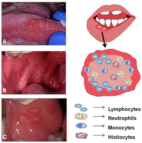
Figure 1. Aphthous ulcers
Notes:
A. Ulcer on the right side of the tongue
B. Ulcer on the posterior buccal mucosa
C. Four ulcers on the anterior buccal mucosa
5. LABORATORY AND DIAGNOSTIC EVALUATION
Laboratory investigations are mainly performed for differential diagnosis and to identify possible underlying causes.
Investigations may include:
Complete blood count
Erythrocyte sedimentation rate
Assessment for nutritional deficiencies, including vitamin B12, folate, and iron
Direct microscopic examination for fungal infection
Cytological examination of vesicular fluid when indicated
HIV testing
Histopathological examination in severe or atypical cases to exclude other mucosal disorders
6. TREATMENT
General Principles
The main goals of treatment are to:
Relieve pain
Promote ulcer healing
Reduce the frequency and severity of recurrent episodes
Maintain good oral hygiene
Avoid factors that may trigger or aggravate lesions
Topical Treatment
Topical corticosteroids are commonly used two to three times daily. Early application at the onset of symptoms can significantly reduce pain and shorten healing time.
Although topical corticosteroids do not completely prevent recurrence, the frequency of recurrent episodes may decrease with treatment.
Other topical treatments that may be used alone or in combination with corticosteroids include:
Topical tetracycline
Sucralfate suspension as a mouthwash four times daily
Hyaluronic acid 0.2% gel or mouthwash
Amlexanox 5% oral paste applied to early lesions four times daily. Adverse effects may include tingling, a cooling sensation at the application site, and a metallic taste.
Laser Therapy
Local helium-neon laser therapy may be applied directly to aphthous ulcers.
Systemic Treatment
First-line systemic treatment
For patients who do not respond adequately to topical therapy alone, a short course of systemic corticosteroids may be considered.
An oral corticosteroid equivalent to prednisone at a dose of 20–40 mg/day may be administered for 4–7 days.
Second-line systemic treatment
For persistent, frequently recurrent disease that cannot be adequately controlled with a short course of systemic corticosteroids combined with topical treatment, additional systemic agents may be considered.
Colchicine: Initial oral dose of 0.6 mg/day for one week. If tolerated, the dose may be increased to 0.6 mg twice daily for at least one month.
Dapsone: Initial dose of 25–50 mg/day, with a maximum dose of 150 mg/day, generally continued for at least one month.
Colchicine and dapsone may also be used in combination, which may provide greater clinical efficacy than either medication alone.
Other Systemic Treatments
Other therapeutic options include:
Thalidomide: 50–100 mg/day, followed by maintenance therapy at 25 mg/day.
Montelukast: A leukotriene receptor antagonist administered at 10 mg/day for one month, followed by 10 mg every other day.
Apremilast: An oral phosphodiesterase inhibitor that suppresses the production of pro-inflammatory cytokines. The recommended regimen is 30 mg twice daily for 2–6 weeks.
Pentoxifylline: 400 mg three times daily.
Supportive Treatment and Pain Management
Secondary infections should be treated if present.
Vitamin and nutritional supplementation may be considered when deficiencies are identified. Vitamin B12 supplementation may be administered for six months.
Topical anesthetics may provide temporary relief, particularly when used before meals and oral hygiene procedures.
Options include:
Viscous lidocaine 2%: A first-line topical anesthetic that may be applied directly to lesions or used as a mouth rinse and then expectorated.
Liquid diphenhydramine: 12.5 mg/5 mL; 5 mL may be used as a mouth rinse.
Dyclonine lozenges: Allowed to dissolve slowly in the mouth.
Aluminum hydroxide, magnesium hydroxide, and simethicone suspension: 5–10 mL used as a mouth rinse and then expectorated.
Attapulgite suspension: 600–750 mg/15 mL; 5–10 mL used as a mouth rinse and then expectorated.
7. PREVENTION
Preventive strategies focus on minimizing known triggers, maintaining good oral hygiene, ensuring adequate nutrition, reducing psychological stress, and identifying and correcting underlying nutritional deficiencies or systemic conditions when present.
REFERENCES
Ministry of Health of Vietnam. Decision No. 4416/QĐ-BYT, 2023. Guidelines for the Diagnosis and Treatment of Dermatological Diseases. Ministry of Health.
Plewa MC, Chatterjee K. Recurrent Aphthous Stomatitis. StatPearls [Internet]. 2025.
Cappello F, Rappa F, Canepa F, et al. Probiotics can cure oral aphthous-like ulcers in inflammatory bowel disease patients: a review of the literature and a working hypothesis. Int J Mol Sci. 2019;20(20):5026. doi:10.3390/ijms20205026.
Kim Ngoc Son, MSc
Nguyen Thanh Tam, MSc
